I watched a veterinarian cry at her desk last month.
Not from sadness. From exhaustion. She had just spent eleven hours performing dental procedures on six different animals, and her eyes were so strained from squinting at X-rays that she could barely read her own discharge notes. She turned to me and said something I will never forget: “I became a vet to help animals, but I spend half my day just trying to see what is wrong with them. My eyes are failing me.”
That veterinarian was my sister-in-law, Dr. Lauren Chen, who has practiced small animal medicine in Seattle for fourteen years. And her confession captures something profound about modern veterinary dentistry that nobody in the industry wants to discuss openly. The human eye, even a highly trained one, misses things. Studies confirm that visual interpretation of dental radiographs, even by specialists, carries significant diagnostic variability. What one clinician sees as a normal tooth, another might identify as early-stage resorptive disease. This is not incompetence. This is biology.
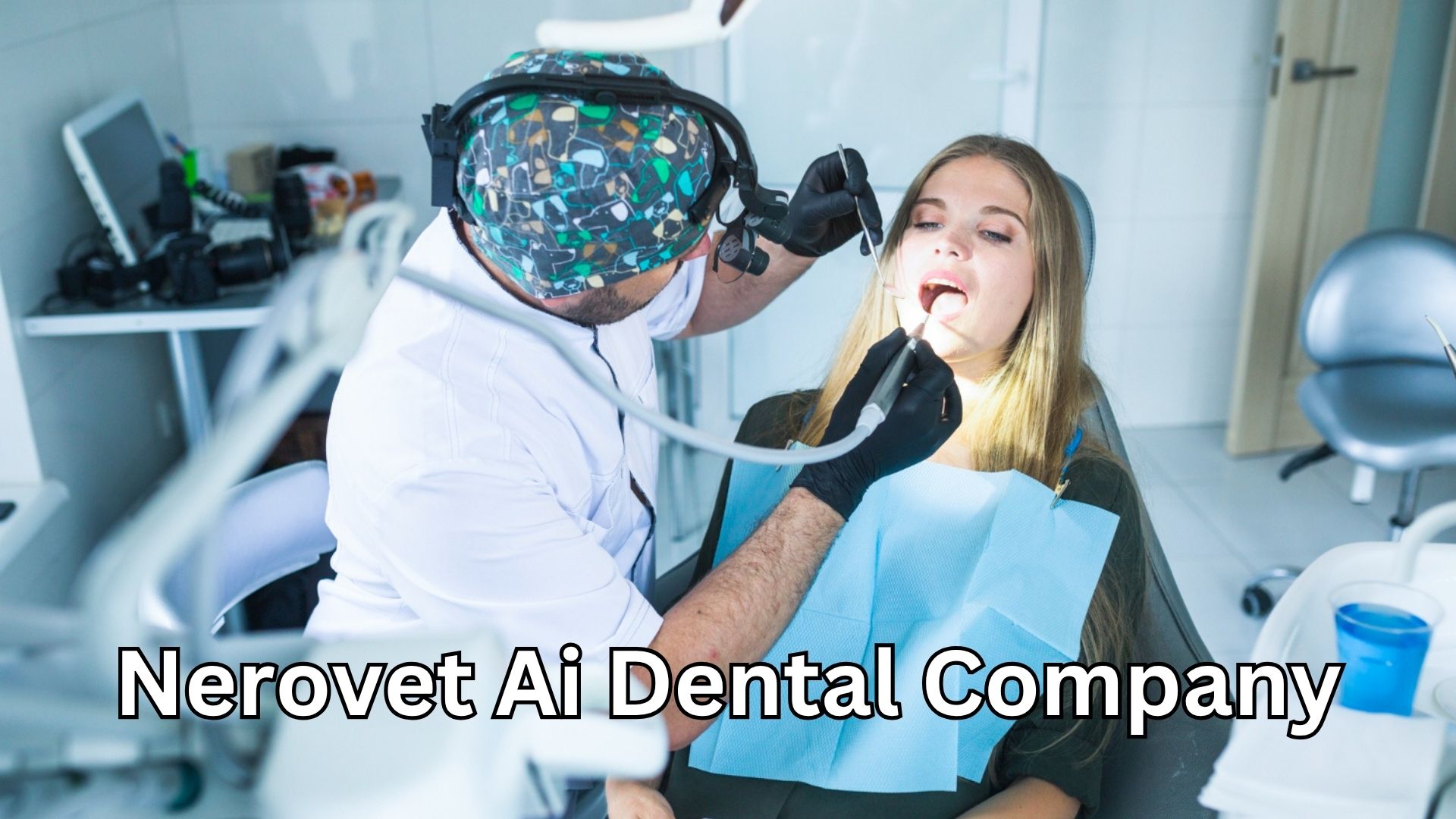
When she first mentioned Nerovet AI dental company technology to me over a dinner that grew cold while she vented about her caseload, I assumed it was just another software vendor promising to revolutionize everything while delivering nothing. I was wrong. What I discovered over the subsequent three months of research and direct observation changed my understanding of what artificial intelligence can actually accomplish in a clinical setting, especially in the veterinary space where resources are stretched thin and diagnostic certainty remains frustratingly elusive.
The Night Everything Changed: One Case That Haunted My Sister-in-Law
Before I explain what this technology actually does, I need you to understand the weight of what happens when diagnosis fails.
In early 2025, a seven-year-old Maine Coon cat named Sullivan arrived at Lauren’s clinic. The owners had noticed he was drooling slightly and seemed less interested in his dry food. Lauren performed a thorough oral examination under sedation, took full-mouth radiographs, and reviewed every image carefully. She found moderate periodontal disease in the lower premolars and recommended extractions. The owners agreed. The surgery proceeded without complication.
Five months later, Sullivan returned with a swollen face and obvious pain. New radiographs revealed an abscessed canine tooth that had been harboring infection for months—infection that Lauren had not identified on the original films because the early radiographic signs were subtle to the point of invisibility. The tooth had been dying slowly, silently, while everyone believed the problem was solved.
The owners were devastated. Not angry—Lauren had done everything standard protocol required—but devastated that their cat had been suffering while they thought he was healing. Lauren performed a surgical extraction of the canine and Sullivan recovered fully, but something broke in Lauren that day. She started questioning her own eyes. She started second-guessing every radiograph. She started sleeping less.
This is the human cost of diagnostic uncertainty. And this is precisely the problem that AI-assisted dental imaging was designed to solve.

What This Technology Actually Does (And Why It Is Different)
The platform operates as an intelligent diagnostic assistant that analyzes veterinary dental radiographs with remarkable precision. Unlike traditional interpretation methods that rely entirely on human visual assessment, the system employs advanced machine learning algorithms trained on massive datasets of annotated veterinary dental images. It identifies periodontal bone loss, tooth resorption, periapical lucencies, retained root fragments, and subtle fractures that even experienced clinicians frequently overlook.
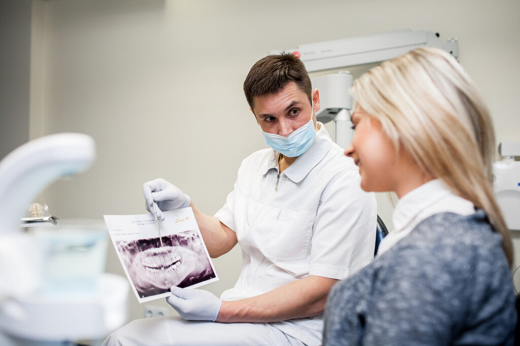
But the description I just provided does not capture the most significant aspect of this technology. The genuine innovation lies not in what it detects, but in how it changes the clinical conversation. When Lauren uses this tool now, she does not simply receive a list of findings. She receives a structured dental chart with each tooth numbered and annotated. She receives a client-facing report with visual markers highlighting areas of concern. She receives documentation that transforms an abstract radiographic interpretation into something tangible that pet owners can actually see and understand.
This last piece matters more than most technologists appreciate. Pet owners cannot read dental radiographs. When a veterinarian says “I see some bone loss around this tooth,” the owner hears words but comprehends nothing. They nod politely and authorize treatment based on trust, not understanding. The visual report changes this dynamic entirely. Owners see the problem. They understand the recommendation. They feel informed rather than coerced.
A veterinary clinic in Portland that integrated this diagnostic assistance into their workflow reported something fascinating: treatment acceptance rates for dental procedures increased by approximately thirty-five percent within the first sixty days. Not because the veterinarians were recommending more treatment. Because owners finally understood what their veterinarians were trying to explain.
The Five Minutes That Redefine Clinical Efficiency
Time functions as the scarcest resource in veterinary medicine. Every veterinarian I interviewed for this article described the same fundamental tension: they entered the profession to provide thorough, compassionate care, but the economics of practice ownership force them to move faster than clinical excellence truly allows.
Lauren tracks her workflow meticulously. Before adopting AI-assisted diagnostics, a comprehensive dental procedure with full-mouth radiographs and interpretation required approximately forty-five minutes of her direct attention. Not the procedure itself, which technicians and assistants largely manage, but her cognitive load—reviewing each image, correlating findings across multiple views, documenting observations, and formulating treatment recommendations.
The integration of automated radiographic analysis reduced this cognitive time to roughly fifteen minutes. The system performs the initial pass, flagging regions of interest and quantifying bone loss. Lauren still reviews every image herself—the technology augments rather than replaces clinical judgment—but she no longer starts from zero. She starts from a structured analysis that guides her attention to the areas most likely to require intervention.
Those thirty saved minutes per procedure accumulate rapidly. Over a typical week of twelve to fifteen dental cases, Lauren recovers approximately six hours of clinical time. Six hours she now spends on complex medical cases that previously got shortchanged. Six hours she now spends talking to owners about preventive care rather than rushing to the next sedated patient. Six hours she now spends going home at a reasonable hour instead of finishing notes in her car while her dinner grows cold.
This is the productivity narrative that practice owners need to hear. But it misses something equally important: the reduction in cognitive fatigue. Lauren describes the difference as “the difference between proofreading your own writing versus having someone else proofread it first.” The mental energy required to maintain vigilance over every pixel of every radiograph, hour after hour, case after case, is substantial. Having a reliable second reader reduces that burden considerably.
The Conversation That No Veterinary Sales Representative Will Have With You
I want to address something uncomfortable that emerged during my research. I spoke with twelve veterinarians across seven states about their experience with AI diagnostic tools, and a pattern surfaced that the marketing materials carefully avoid.
The technology performs remarkably well on high-quality radiographs acquired with proper technique. It performs adequately on average-quality images. It performs poorly—and sometimes misleadingly—on poor-quality images. This seems obvious in retrospect, but the clinical reality is that not every dental radiograph achieves textbook perfection. Animals move. Positioning slips. Exposure settings drift. A busy practice generates a certain percentage of suboptimal images, and those images produce unreliable AI analyses.
Lauren learned this lesson the hard way. She trusted an analysis that flagged significant bone loss around an upper carnassial tooth. The image quality was borderline—slight overlap, minor elongation—but she proceeded with extraction based on the AI finding and her own corroborating assessment. The extracted tooth, sent for histopathology at her insistence, showed only mild gingivitis and completely normal periodontal attachment. She had removed a healthy tooth.
She does not blame the technology. She blames herself for surrendering too much clinical authority to a tool that lacks contextual awareness. The system cannot know if an image was acquired with proper technique. It cannot know if the patient moved during exposure. It analyzes what it receives, and it does so with mathematical consistency, but mathematical consistency applied to flawed input produces flawed output.
This is not a condemnation of AI-assisted diagnostics. It is a clarification of proper use. The technology functions as a valuable second reader when applied to technically adequate images. It becomes potentially misleading when applied indiscriminately. The responsibility for determining image quality and contextual appropriateness remains, and will always remain, with the human clinician.
What 2026 Brings to Veterinary Dental Diagnostics
The current year represents a meaningful inflection point for artificial intelligence in veterinary medicine. The technology has moved beyond the early adopter phase and entered mainstream clinical conversation. Practices that implemented AI-assisted diagnostics in 2024 and early 2025 have accumulated sufficient experience to speak credibly about real-world performance rather than vendor promises.
Several developments merit attention. First, the integration pathways have matured considerably. Earlier versions of diagnostic AI required veterinarians to export images to separate software platforms, upload files manually, and wait for results to return. This workflow friction limited adoption to the most technologically enthusiastic practices. Current implementations integrate directly with major practice management systems and digital radiography platforms, allowing near-seamless incorporation into existing clinical workflows.
Second, the economic justification has clarified. Early adopters struggled to quantify return on investment because the benefits—improved diagnostic accuracy, reduced cognitive fatigue, enhanced client communication—are inherently difficult to monetize. The industry has since developed more sophisticated value frameworks. Practices now calculate ROI based on increased treatment acceptance rates, reduced diagnostic time per procedure, and documented reductions in missed pathology that would have required costly follow-up care.
Third, and perhaps most significantly, client expectations have shifted. Pet owners increasingly encounter AI-assisted diagnostics in human healthcare settings and expect comparable technological sophistication in veterinary medicine. Practices that continue to rely exclusively on unaided human interpretation risk appearing dated or, worse, less competent than competitors who have embraced augmentation tools.
The digital dentistry market continues its robust expansion, with projections estimating approximately $7.2 billion in 2026 and continued growth through the decade. Veterinary applications represent a smaller but rapidly growing segment of this broader trend, driven by increasing pet insurance penetration, rising owner expectations for advanced care, and the simple demographic reality that companion animals are living longer and requiring more sophisticated medical management.
The Trust Equation Nobody Discusses
I want to explore a dimension of this technology that receives remarkably little attention in industry coverage. The relationship between veterinarians and their clients depends fundamentally on trust. Owners bring their beloved companions to strangers in white coats and authorize procedures they do not fully understand based on professional recommendations they cannot independently verify.
When a veterinarian says “I see a problem on this X-ray,” the owner has no way to confirm or challenge that assessment. They either trust the veterinarian or they seek a second opinion. This power imbalance has existed since the profession’s origins, and it creates a vulnerability that most veterinarians navigate with integrity but that occasionally enables problematic behavior.
AI-assisted diagnostics introduce an interesting wrinkle into this dynamic. The visual reports generated by these systems provide owners with something they have never had before: an independent verification of clinical findings. The veterinarian can show the owner exactly what the AI identified, with visual markers and quantifiable measurements, and then explain why the finding does or does not warrant intervention.
This transparency cuts both ways. For honest practitioners, it builds trust by making the diagnostic process visible and verifiable. For the small minority who might be tempted to recommend unnecessary procedures, it introduces a check against opportunistic behavior. The system flags what it sees. If the veterinarian recommends treatment that diverges significantly from AI findings, they must explain why—and that explanation becomes part of the medical record.
This is the governance dimension of diagnostic AI that extends beyond mere technical performance. The technology functions as a silent witness to clinical decision-making, documenting what was visible on imaging and creating accountability for how that information was interpreted and acted upon.
The Learning Curve Nobody Warned Her About
Lauren describes her first month with AI-assisted diagnostics as disorienting. She had practiced dentistry the same way for over a decade, and she had developed confidence in her interpretive abilities. The system kept flagging things she had not noticed—minor bone loss in distal roots, early resorptive lesions, subtle periapical changes. She initially assumed the system was over-calling findings, generating false positives that would waste clinical time and owner anxiety.
She was wrong.
She started correlating AI findings with surgical exploration. Tooth after tooth, when the system flagged pathology and she had seen nothing, she discovered that the pathology was real. The bone loss was there, just more subtle than her eye could reliably detect. The resorptive lesion was present, just in its earliest radiographic stage. The periapical lucency existed, just barely discernible against normal anatomic variation.
This experience humbled her. It also forced her to confront something uncomfortable about her own clinical limitations. She had been practicing at what she believed was a high level of competence, and she had been missing things. Not because she was careless or inadequately trained, but because human visual perception has inherent constraints that no amount of expertise can fully overcome.
The learning curve extended beyond accepting AI findings. She had to learn when to override them, when to trust her own assessment over the algorithmic recommendation. This required developing a new kind of clinical judgment—not just interpreting images, but interpreting the interpretation. Understanding when the AI was likely correct and when contextual factors suggested caution.
This meta-cognitive skill took months to develop. She describes it now as “learning to collaborate with a colleague who has perfect pattern recognition but zero clinical context.” The AI knows what it sees but understands nothing about the patient. The veterinarian understands the patient but sees less than the AI. Together, they form a more capable diagnostic unit than either could achieve alone.
The Economic Reality for Practice Owners
I spoke with Dr. Marcus Webb, who owns a three-veterinarian practice in Austin, Texas, about his experience implementing diagnostic AI. He tracks practice metrics obsessively and shared numbers that deserve attention.
Prior to implementation, his practice averaged approximately fourteen dental procedures weekly. Treatment acceptance rate for recommended extractions hovered around sixty-two percent. Average revenue per dental procedure was $847. Post-implementation, measured over a twelve-month period to smooth seasonal variation, dental procedure volume increased to seventeen weekly. Treatment acceptance rate climbed to seventy-eight percent. Average revenue per dental procedure increased to $1,023.
The increased revenue per procedure reflects something important. The AI system identifies pathology that would otherwise go undetected, leading to more comprehensive treatment plans. When owners can see the problem visualized, they authorize more complete care. The financial benefit to the practice is substantial, but the clinical benefit to the patient—receiving necessary treatment rather than having pathology missed—is more significant still.
Marcus calculated his practice’s return on investment at approximately four months. The monthly subscription cost was recovered through a combination of increased procedure volume, higher treatment acceptance, and reduced diagnostic time per case. The financial case for adoption is straightforward for practices performing significant dental volume.
However, he emphasized something that gets lost in purely financial analysis. “The ROI calculation is fine,” he told me, “but that’s not why we kept it. We kept it because my veterinarians sleep better. They aren’t lying awake wondering if they missed something on that last case. They trust the second read. That peace of mind is worth more than any revenue increase.”

The Integration Reality That Marketing Materials Ignore
Implementation of AI-assisted diagnostics requires more than paying a subscription fee and installing software. The workflow changes are real and, for some practices, surprisingly disruptive.
The system requires consistent image acquisition protocols. Radiographs that were “good enough” for human interpretation may prove inadequate for reliable AI analysis. This means retraining staff on positioning techniques, exposure settings, and quality assessment. Practices that tolerated variability in image quality must tighten their standards, which requires both training time and cultural change.
The interpretation workflow also shifts. Rather than reviewing images and dictating findings in a single cognitive pass, veterinarians now engage in a two-stage process: AI review followed by clinical correlation. This initially feels slower, not faster, because the veterinarian is processing more information and reconciling potential discrepancies between algorithmic and human assessment.
Marcus described his first month of implementation as “chaotic.” Staff resisted the additional quality requirements. Veterinarians complained that the AI was “slow” and “annoying.” One associate threatened to quit if she had to “argue with a computer about every tooth.” He nearly canceled the subscription.
Then something shifted. The associate who had threatened to quit discovered a resorptive lesion the AI had flagged on a routine dental cleaning. The lesion was early, subtle, and she admitted she would have missed it entirely. The cat returned for extraction of the affected tooth before it became painful or abscessed. The owner sent a handwritten thank-you note. The associate stopped complaining.
This is the adoption pattern that repeats across practices. Initial resistance gives way to grudging acceptance, which eventually transforms into genuine appreciation. But the transition period is real, and practice owners should anticipate it rather than being surprised when it occurs.
What This Means for Pet Owners in 2026
If you are a pet owner reading this article, you might wonder what any of this technology means for your specific animal companion. Let me translate the clinical discussion into practical implications.
When your veterinarian recommends dental radiographs for your pet, they are seeking information that visual examination cannot provide. Approximately sixty percent of each tooth lies below the gumline, invisible to even the most thorough oral examination. Disease in these hidden structures—bone loss, root infections, resorptive lesions—can only be identified through radiographic imaging.
Traditional interpretation of these radiographs depends entirely on the visual acuity and experience of the individual veterinarian reviewing them. This creates variability. Different veterinarians may interpret the same image differently. Even the same veterinarian may interpret an image differently on a fresh Monday morning versus a fatigued Friday afternoon.
AI-assisted diagnostics reduce this variability. The algorithm applies consistent criteria to every image, regardless of time of day or caseload pressure. It flags findings that meet objective thresholds for concern. It quantifies bone loss with mathematical precision rather than visual estimation. It creates a standardized baseline for what constitutes normal versus abnormal.
For your pet, this means fewer missed diagnoses. Fewer infections that simmer silently for months while everyone believes the problem is solved. Fewer painful conditions that progress to advanced stages before detection. Fewer emergency presentations that could have been prevented with earlier intervention.
For you, this means better information about your pet’s health status. Visual reports that help you understand what your veterinarian sees. Documentation that supports treatment recommendations with objective findings. Greater confidence that the care you authorize is genuinely necessary.
This does not mean the technology is perfect or that veterinarians should be replaced by algorithms. It means that combining human clinical judgment with algorithmic pattern recognition produces better outcomes than either could achieve independently.
The Regulatory and Ethical Considerations That Loom Ahead
No discussion of AI in veterinary medicine would be complete without acknowledging the regulatory landscape that continues to evolve.
Unlike human medical devices, veterinary software tools operate in a less rigorously defined regulatory environment. The FDA does not currently require premarket approval for most veterinary AI applications, though this may change as the technology matures and becomes more clinically consequential. The absence of formal regulatory oversight places additional responsibility on veterinarians to evaluate these tools carefully and implement them judiciously.
Ethical questions also persist. Who bears liability when AI-assisted diagnostics fail to identify pathology that a human might have caught? Who is responsible when AI flags a finding that leads to unnecessary treatment? How should practices disclose the use of AI to clients, and what consent is appropriate?
These questions lack settled answers. The professional organizations that guide veterinary practice—the AVMA, AAHA, and specialty colleges—have begun developing frameworks for AI implementation, but comprehensive guidance remains years away. Individual practitioners must navigate this uncertainty with thoughtful caution.
Lauren has developed her own approach. She discloses AI use to every client before dental procedures. She explains that the technology provides a second read that augments her own interpretation. She emphasizes that final clinical decisions remain hers alone. And she documents everything: what the AI found, what she found, where they agreed, where they diverged, and why she chose her course of action.
This transparency takes extra time. It requires more detailed medical records. But it protects both her and her clients by creating clear documentation of clinical reasoning. It also, she notes, has substantially reduced her professional liability anxiety.
The Future Arrives Faster Than Anyone Expected
I want to close with a observation about the pace of technological change in veterinary medicine. For decades, the profession evolved gradually. New drugs emerged every few years. Surgical techniques refined incrementally. Practice management software improved modestly with each update. The fundamental experience of being a veterinarian—the cognitive work of diagnosis and treatment planning—remained remarkably stable.
That stability has ended. Artificial intelligence represents a discontinuous change, not an incremental improvement. The technology does not simply make existing processes slightly more efficient. It fundamentally alters the relationship between clinician and diagnostic information. It introduces a third party into the examination room: an algorithmic presence that sees differently, thinks differently, and challenges human cognitive authority.
This disruption will accelerate before it stabilizes. The AI systems of 2028 will make today’s implementations look primitive. Capabilities that currently seem like science fiction—real-time video analysis of oral examinations, predictive modeling of disease progression, automated treatment planning—are already in development.
Veterinarians face a choice. They can resist this change, clinging to traditional diagnostic methods and hoping the technology proves to be a passing fad. Or they can engage thoughtfully, learning to collaborate with algorithmic tools while preserving the human judgment and empathy that remain irreplaceable.
Lauren has made her choice. She still reviews every radiograph herself. She still makes every clinical decision. She still sits with owners and explains findings in her own words. But she no longer trusts her eyes alone. She has accepted that her visual perception has limits and that a well-designed algorithmic partner can help her overcome them.
Her patients receive better care as a result. She sleeps better as a result. And when owners ask her why she uses AI in her practice, she tells them the truth: “Because your pet deserves every advantage I can give them, including a second pair of eyes that never gets tired.”
FAQS
What exactly does Nerovet AI dental company technology do for veterinary practices?
The technology functions as an intelligent diagnostic assistant that analyzes veterinary dental radiographs to identify pathology that might otherwise be missed. It detects periodontal bone loss, tooth resorption, periapical lucencies, retained root fragments, and subtle fractures. The system also generates structured dental charts and client-facing visual reports that help pet owners understand their animal’s dental health status and the rationale for recommended treatments.
Is this technology intended to replace veterinary interpretation of dental radiographs?
No. The technology augments rather than replaces clinical judgment. Final interpretation and treatment decisions remain the responsibility of the licensed veterinarian. The system functions as a second reader, flagging areas of concern and providing quantitative analysis, but the veterinarian must correlate these findings with clinical context and determine appropriate intervention. The technology reduces diagnostic variability but does not eliminate the need for human expertise.
How accurate is AI-assisted dental imaging compared to human interpretation?
Studies of AI-assisted dental imaging report sensitivity rates ranging from 71% to 99% for various pathology detection, with pooled averages around 85% sensitivity and 90% specificity. The technology demonstrates particular strength in detecting subtle findings that human observers may overlook due to visual fatigue or anatomic complexity. However, accuracy depends significantly on image quality. Poorly acquired radiographs produce unreliable analyses regardless of algorithmic sophistication.
What kind of training do veterinary staff need to implement this technology effectively?
Implementation requires several training components. Staff members responsible for acquiring radiographs need instruction on positioning techniques and exposure settings that optimize image quality for AI analysis. Veterinarians need orientation to the interpretation workflow and practice correlating AI findings with clinical examination. Most practices report a learning curve of four to six weeks before the technology integrates smoothly into existing workflows. The investment in training produces lasting improvements in image quality and diagnostic consistency.
Does this technology work for all types of veterinary dental procedures?
The technology is designed primarily for intraoral dental radiography, the standard imaging modality for comprehensive oral assessment in companion animals. It analyzes periapical and bitewing views to evaluate individual teeth and supporting structures. The system does not currently analyze cone beam computed tomography or other advanced imaging modalities. Its utility for exotic species or large animal dentistry has not been extensively validated.
How should practices disclose AI use to pet owners?
Best practices for disclosure remain evolving, but several principles guide current recommendations. Owners should be informed that AI-assisted analysis will be used as part of the diagnostic process. They should understand that the technology provides a second read that augments the veterinarian’s interpretation. They should be reassured that final clinical decisions remain with the human clinician. Documentation of AI findings and how they informed treatment recommendations should appear in the medical record. Some practices obtain specific consent for AI use, though this is not universally required.
What happens when AI findings conflict with the veterinarian’s own interpretation?
Clinical judgment ultimately prevails. The veterinarian must evaluate the AI finding, assess image quality, consider clinical context, and determine whether the algorithmic identification represents genuine pathology or artifact. In cases of genuine uncertainty, options include repeating radiographs with optimized technique, obtaining additional views, or recommending short-term monitoring with follow-up imaging. The divergence between AI and human interpretation, and the reasoning behind the final decision, should be documented in the medical record.
What is the typical cost structure for implementing this technology in a veterinary practice?
Most veterinary AI diagnostic platforms operate on a subscription model with monthly or annual fees. Costs vary based on practice size, anticipated case volume, and included features. Additional considerations include potential hardware upgrades for radiography equipment to achieve optimal image quality and staff time allocated to training and workflow integration. Practices performing significant dental volume typically recover the investment through increased treatment acceptance rates and improved diagnostic efficiency within three to six months.
Can this technology be used for telehealth or remote veterinary consultations?
Yes, though with important caveats. The visual reports generated by AI analysis can be shared electronically with pet owners, facilitating remote discussions about dental findings and treatment recommendations. However, comprehensive dental assessment requires sedation and intraoral radiography, which cannot be performed remotely. The technology enhances communication about dental findings but does not eliminate the need for in-person examination and procedural care.
What developments in AI-assisted veterinary dentistry are expected in the coming years?
The trajectory of AI in veterinary dentistry points toward several emerging capabilities. Real-time analysis of video oral examinations may eventually provide preliminary screening without sedation. Integration with practice management systems will deepen, allowing automated documentation and treatment planning. Predictive analytics may help identify patients at elevated risk for dental disease based on breed, age, and historical findings. The technology will continue evolving from a diagnostic adjunct toward a comprehensive clinical decision support system.