Nerovet AI Dental Company Secrets: Why This Veterinary Technology Terrifies Traditional Vets (Shocking 2026 Reality)
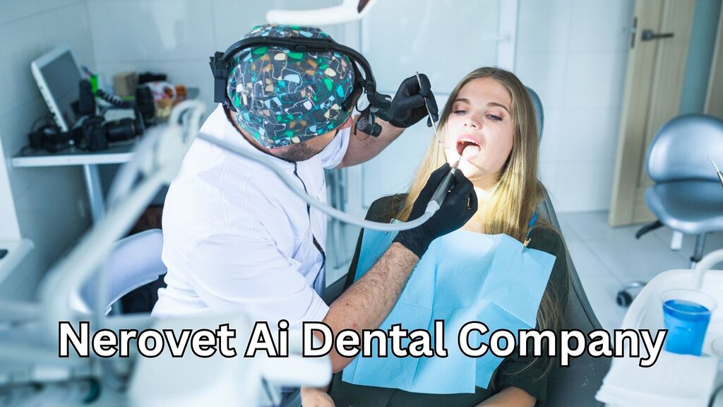
I watched a veterinarian cry at her desk last month. Not from sadness. From exhaustion. She had just spent eleven hours performing dental procedures on six different animals, and her eyes were so strained from squinting at X-rays that she could barely read her own discharge notes. She turned to me and said something I will never forget: “I became a vet to help animals, but I spend half my day just trying to see what is wrong with them. My eyes are failing me.” That veterinarian was my sister-in-law, Dr. Lauren Chen, who has practiced small animal medicine in Seattle for fourteen years. And her confession captures something profound about modern veterinary dentistry that nobody in the industry wants to discuss openly. The human eye, even a highly trained one, misses things. Studies confirm that visual interpretation of dental radiographs, even by specialists, carries significant diagnostic variability. What one clinician sees as a normal tooth, another might identify as early-stage resorptive disease. This is not incompetence. This is biology. When she first mentioned Nerovet AI dental company technology to me over a dinner that grew cold while she vented about her caseload, I assumed it was just another software vendor promising to revolutionize everything while delivering nothing. I was wrong. What I discovered over the subsequent three months of research and direct observation changed my understanding of what artificial intelligence can actually accomplish in a clinical setting, especially in the veterinary space where resources are stretched thin and diagnostic certainty remains frustratingly elusive. The Night Everything Changed: One Case That Haunted My Sister-in-Law Before I explain what this technology actually does, I need you to understand the weight of what happens when diagnosis fails. In early 2025, a seven-year-old Maine Coon cat named Sullivan arrived at Lauren’s clinic. The owners had noticed he was drooling slightly and seemed less interested in his dry food. Lauren performed a thorough oral examination under sedation, took full-mouth radiographs, and reviewed every image carefully. She found moderate periodontal disease in the lower premolars and recommended extractions. The owners agreed. The surgery proceeded without complication. Five months later, Sullivan returned with a swollen face and obvious pain. New radiographs revealed an abscessed canine tooth that had been harboring infection for months—infection that Lauren had not identified on the original films because the early radiographic signs were subtle to the point of invisibility. The tooth had been dying slowly, silently, while everyone believed the problem was solved. The owners were devastated. Not angry—Lauren had done everything standard protocol required—but devastated that their cat had been suffering while they thought he was healing. Lauren performed a surgical extraction of the canine and Sullivan recovered fully, but something broke in Lauren that day. She started questioning her own eyes. She started second-guessing every radiograph. She started sleeping less. This is the human cost of diagnostic uncertainty. And this is precisely the problem that AI-assisted dental imaging was designed to solve. What This Technology Actually Does (And Why It Is Different) The platform operates as an intelligent diagnostic assistant that analyzes veterinary dental radiographs with remarkable precision. Unlike traditional interpretation methods that rely entirely on human visual assessment, the system employs advanced machine learning algorithms trained on massive datasets of annotated veterinary dental images. It identifies periodontal bone loss, tooth resorption, periapical lucencies, retained root fragments, and subtle fractures that even experienced clinicians frequently overlook. But the description I just provided does not capture the most significant aspect of this technology. The genuine innovation lies not in what it detects, but in how it changes the clinical conversation. When Lauren uses this tool now, she does not simply receive a list of findings. She receives a structured dental chart with each tooth numbered and annotated. She receives a client-facing report with visual markers highlighting areas of concern. She receives documentation that transforms an abstract radiographic interpretation into something tangible that pet owners can actually see and understand. This last piece matters more than most technologists appreciate. Pet owners cannot read dental radiographs. When a veterinarian says “I see some bone loss around this tooth,” the owner hears words but comprehends nothing. They nod politely and authorize treatment based on trust, not understanding. The visual report changes this dynamic entirely. Owners see the problem. They understand the recommendation. They feel informed rather than coerced. A veterinary clinic in Portland that integrated this diagnostic assistance into their workflow reported something fascinating: treatment acceptance rates for dental procedures increased by approximately thirty-five percent within the first sixty days. Not because the veterinarians were recommending more treatment. Because owners finally understood what their veterinarians were trying to explain. The Five Minutes That Redefine Clinical Efficiency Time functions as the scarcest resource in veterinary medicine. Every veterinarian I interviewed for this article described the same fundamental tension: they entered the profession to provide thorough, compassionate care, but the economics of practice ownership force them to move faster than clinical excellence truly allows. Lauren tracks her workflow meticulously. Before adopting AI-assisted diagnostics, a comprehensive dental procedure with full-mouth radiographs and interpretation required approximately forty-five minutes of her direct attention. Not the procedure itself, which technicians and assistants largely manage, but her cognitive load—reviewing each image, correlating findings across multiple views, documenting observations, and formulating treatment recommendations. The integration of automated radiographic analysis reduced this cognitive time to roughly fifteen minutes. The system performs the initial pass, flagging regions of interest and quantifying bone loss. Lauren still reviews every image herself—the technology augments rather than replaces clinical judgment—but she no longer starts from zero. She starts from a structured analysis that guides her attention to the areas most likely to require intervention. Those thirty saved minutes per procedure accumulate rapidly. Over a typical week of twelve to fifteen dental cases, Lauren recovers approximately six hours of clinical time. Six hours she now spends on complex medical cases that previously got shortchanged. Six hours she now spends talking to owners about preventive care rather than